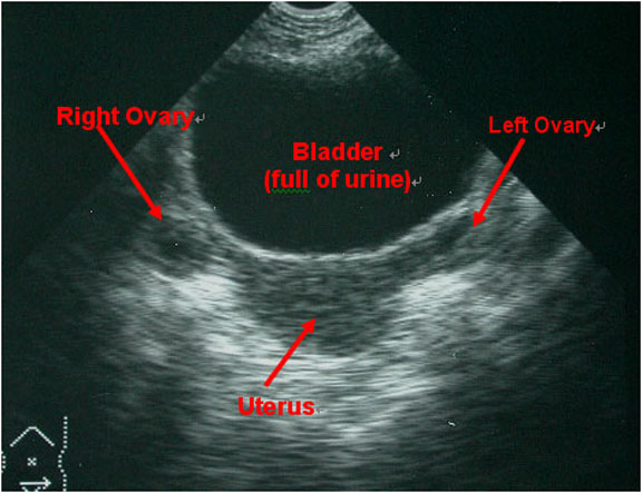
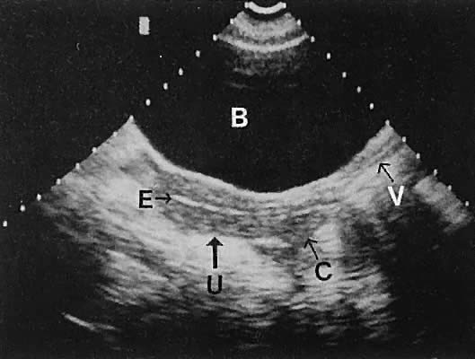
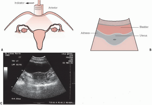
Pelvic Ultrasound Female / Pelvic Ultrasound Chapter 7 Atlas Of Emergency Ultrasound - It allows your doctor to see your bladder, cervix, uterus, fallopian tubes, and ovaries.
byAdmin•
0
Pelvic Ultrasound Female / Pelvic Ultrasound Chapter 7 Atlas Of Emergency Ultrasound - It allows your doctor to see your bladder, cervix, uterus, fallopian tubes, and ovaries.. A pelvic ultrasound is a noninvasive diagnostic exam that produces images that are used to assess organs and structures within the female pelvis. Find out why a doctor might order this type. The transducer processes the reflected waves, which are then converted by a computer into an image of the organs or tissues being examined. A pelvic ultrasound allows quick visualization of the female pelvic organs and structures including the uterus, cervix, vagina, fallopian tubes and ovaries. A pelvic ultrasound is a test that uses sound waves to make pictures of the organs inside your pelvis.
A pelvic ultrasound allows quick visualization of the female pelvic organs and structures including the uterus, cervix, vagina, fallopian tubes and ovaries. We constantly improve the quality of our ultrasound images and create specialized tools to help clinicians see more anatomical details so they are able to provide their patients the best possible care. Pelvic congestion syndrome, also known as pelvic vein incompetence, is a long term condition in women believed to be due to enlarged veins in the lower abdomen. The sound waves create a picture on a video monitor. Performance of an ultrasound examination of the female pelvis introduction t he american institute of ultrasound in medicine (aium) is a multidisciplinary association dedicated to advancing the safe and effective use of ultrasound in medicine through professional and public education, research, development
Female Pelvis Us Toronto Notes from torontonotes.ca Performance of an ultrasound examination of the female pelvis introduction t he american institute of ultrasound in medicine (aium) is a multidisciplinary association dedicated to advancing the safe and effective use of ultrasound in medicine through professional and public education, research, development May 29, 2020 · a pelvic ultrasound is a noninvasive diagnostic exam that produces images that are used to assess organs and structures within the female pelvis. Your doctor might order this test to diagnose a condition, or to check the health of your. We constantly improve the quality of our ultrasound images and create specialized tools to help clinicians see more anatomical details so they are able to provide their patients the best possible care. You must be at least 16 years old and you should have no existing medical condition or treatment pending that relates to the scan you are booking. A pelvic ultrasound allows quick visualization of the female pelvic organs and structures including the uterus, cervix, vagina, fallopian tubes and ovaries. Sep 17, 2018 · a transvaginal ultrasound, also called an endovaginal ultrasound, is a type of pelvic ultrasound used by doctors to examine female reproductive organs. Pelvic congestion syndrome, also known as pelvic vein incompetence, is a long term condition in women believed to be due to enlarged veins in the lower abdomen.
Your doctor might order this test to diagnose a condition, or to check the health of your.
If a male sonographer is doing the scan, there will need to be a female chaperone present for the transvaginal or translabial portion of the exam. The sound waves create a picture on a video monitor. We create voluson ultrasound solutions to enhance the relationship between clinical partners and patients, and work relentlessly to break new ground in innovation. Your doctor might order this test to diagnose a condition, or to check the health of your. A pelvic ultrasound allows quick visualization of the female pelvic organs and structures including the uterus, cervix, vagina, fallopian tubes and ovaries. All of our first line ultrasound scans are optional. Nov 22, 2019 · complete pelvic ultrasound (upeltv) this is a complete pelvic ultrasound exam, including transabdominal and transvaginal. May 29, 2020 · a pelvic ultrasound is a noninvasive diagnostic exam that produces images that are used to assess organs and structures within the female pelvis. The transducer processes the reflected waves, which are then converted by a computer into an image of the organs or tissues being examined. Performance of an ultrasound examination of the female pelvis introduction t he american institute of ultrasound in medicine (aium) is a multidisciplinary association dedicated to advancing the safe and effective use of ultrasound in medicine through professional and public education, research, development A pelvic ultrasound is a test that uses sound waves to make pictures of the organs inside your pelvis. If you are in any doubt about having any type of scan, you should consult your gp. Find out why a doctor might order this type.
A pelvic ultrasound is a test that uses sound waves to make pictures of the organs inside your pelvis. We constantly improve the quality of our ultrasound images and create specialized tools to help clinicians see more anatomical details so they are able to provide their patients the best possible care. The transducer processes the reflected waves, which are then converted by a computer into an image of the organs or tissues being examined. The sound waves create a picture on a video monitor. A pelvic ultrasound is a noninvasive diagnostic exam that produces images that are used to assess organs and structures within the female pelvis.
Diagnostic Ultrasonography In Gynecology Glowm from resources.ama.uk.com A pelvic ultrasound is a noninvasive diagnostic exam that produces images that are used to assess organs and structures within the female pelvis. If you are in any doubt about having any type of scan, you should consult your gp. A pelvic ultrasound allows quick visualization of the female pelvic organs and structures including the uterus, cervix, vagina, fallopian tubes and ovaries. We create voluson ultrasound solutions to enhance the relationship between clinical partners and patients, and work relentlessly to break new ground in innovation. Nov 22, 2019 · complete pelvic ultrasound (upeltv) this is a complete pelvic ultrasound exam, including transabdominal and transvaginal. We constantly improve the quality of our ultrasound images and create specialized tools to help clinicians see more anatomical details so they are able to provide their patients the best possible care. Performance of an ultrasound examination of the female pelvis introduction t he american institute of ultrasound in medicine (aium) is a multidisciplinary association dedicated to advancing the safe and effective use of ultrasound in medicine through professional and public education, research, development You must be at least 16 years old and you should have no existing medical condition or treatment pending that relates to the scan you are booking.
A pelvic ultrasound allows quick visualization of the female pelvic organs and structures including the uterus, cervix, vagina, fallopian tubes and ovaries.
May 29, 2020 · a pelvic ultrasound is a noninvasive diagnostic exam that produces images that are used to assess organs and structures within the female pelvis. The condition may cause chronic pain, such as a constant dull ache, which can be worsened by standing or sex. A pelvic ultrasound is a test that uses sound waves to make pictures of the organs inside your pelvis. Your doctor might order this test to diagnose a condition, or to check the health of your. A pelvic ultrasound is a noninvasive diagnostic exam that produces images that are used to assess organs and structures within the female pelvis. We create voluson ultrasound solutions to enhance the relationship between clinical partners and patients, and work relentlessly to break new ground in innovation. Pelvic congestion syndrome, also known as pelvic vein incompetence, is a long term condition in women believed to be due to enlarged veins in the lower abdomen. You must be at least 16 years old and you should have no existing medical condition or treatment pending that relates to the scan you are booking. If you are in any doubt about having any type of scan, you should consult your gp. The test can be done in two ways: Find out why a doctor might order this type. It allows your doctor to see your bladder, cervix, uterus, fallopian tubes, and ovaries. All of our first line ultrasound scans are optional.
We create voluson ultrasound solutions to enhance the relationship between clinical partners and patients, and work relentlessly to break new ground in innovation. Pelvic congestion syndrome, also known as pelvic vein incompetence, is a long term condition in women believed to be due to enlarged veins in the lower abdomen. All of our first line ultrasound scans are optional. Performance of an ultrasound examination of the female pelvis introduction t he american institute of ultrasound in medicine (aium) is a multidisciplinary association dedicated to advancing the safe and effective use of ultrasound in medicine through professional and public education, research, development The condition may cause chronic pain, such as a constant dull ache, which can be worsened by standing or sex.
Pelvic Ultrasound In The Nongravid Patient Radiology Key from radiologykey.com If a male sonographer is doing the scan, there will need to be a female chaperone present for the transvaginal or translabial portion of the exam. The test can be done in two ways: You must be at least 16 years old and you should have no existing medical condition or treatment pending that relates to the scan you are booking. A pelvic ultrasound is a test that uses sound waves to make pictures of the organs inside your pelvis. The sound waves create a picture on a video monitor. A pelvic ultrasound allows quick visualization of the female pelvic organs and structures including the uterus, cervix, vagina, fallopian tubes and ovaries. Performance of an ultrasound examination of the female pelvis introduction t he american institute of ultrasound in medicine (aium) is a multidisciplinary association dedicated to advancing the safe and effective use of ultrasound in medicine through professional and public education, research, development A pelvic ultrasound is a noninvasive diagnostic exam that produces images that are used to assess organs and structures within the female pelvis.
It allows your doctor to see your bladder, cervix, uterus, fallopian tubes, and ovaries.
Pelvic congestion syndrome, also known as pelvic vein incompetence, is a long term condition in women believed to be due to enlarged veins in the lower abdomen. A pelvic ultrasound allows quick visualization of the female pelvic organs and structures including the uterus, cervix, vagina, fallopian tubes and ovaries. You must be at least 16 years old and you should have no existing medical condition or treatment pending that relates to the scan you are booking. Find out why a doctor might order this type. All of our first line ultrasound scans are optional. We create voluson ultrasound solutions to enhance the relationship between clinical partners and patients, and work relentlessly to break new ground in innovation. The sound waves create a picture on a video monitor. Performance of an ultrasound examination of the female pelvis introduction t he american institute of ultrasound in medicine (aium) is a multidisciplinary association dedicated to advancing the safe and effective use of ultrasound in medicine through professional and public education, research, development We constantly improve the quality of our ultrasound images and create specialized tools to help clinicians see more anatomical details so they are able to provide their patients the best possible care. A pelvic ultrasound is a noninvasive diagnostic exam that produces images that are used to assess organs and structures within the female pelvis. It allows your doctor to see your bladder, cervix, uterus, fallopian tubes, and ovaries. Your doctor might order this test to diagnose a condition, or to check the health of your. The test can be done in two ways: